Almost 3- 5% of the infants worldwide are born with birth defects and genetic abnormalities. Among them, chromosomal abnormalities are present in 1/150 live births. These congenital abnormalities are considered to be the root cause of neonatal deaths. With advancement in technologies, prenatal screening and diagnosis have become a beneficial approach to determine foetal viability for congenital abnormalities and birth defects. Early information about these problems provides option to patients(soon-to-be parents) for their further proceedings. If defect get detected at early gestational period/age, they may choose pregnancy termination or they may choose to pursue the pregnancy, where they seek experienced clinicians who may able to guide them for carrying an affected infant and to care for the child after delivery.
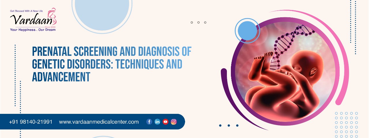
PRENATAL SCREENING AND DIAGNOSIS OF GENETIC DISORDERS: TECHNIQUES AND ADVANCEMENT
Prenatal screening and prenatal diagnosis is a procedure to diagnose problems associated with pregnancy as early as possible. These problems can be anatomical and physiological such as neural tube defects, chromosomal abnormalities and gene mutations that results into genetic disorder or birth defects such as Down’s syndrome, spina bifida, cystic fibrosis and muscular dystrophy and others.
However, prenatal diagnosis and prenatal screening share a common purpose but both the terms means differently. Prenatal screening mainly focuses on finding a problem whereas prenatal diagnosis focuses on pursuing an additional information once a particular problem has been diagnosed.
Indication of prenatal screening and diagnosis:
• Advanced maternal age (AMA).
• Child with chromosomal abnormality.
• Twin or multiple pregnancy.
• Family history of single gene disorder, neural tube defects and other congenital
abnormalities.
• Abnormalities identified in pregnancy.
• History of miscarriages.
• Other high risks such as consanguinity , maternal illness.
• Invasive prenatal screening and diagnosis
Prenatal screening has evolved with traditional invasive methods like
Amniocentesis: – amniocentesis is carried between 15 to 17 weeks of pregnancy under ultrasonographic guidance. About 15 ml of amniotic fluid aspirated for cell culture followed by chromosomal analysis. Metaphase chromosomes are analysed for structural and numerical abnormalities. Uncultured amniotic fluid is used to estimate level of Alpha Feto Protein (AFP). Raised levels of AFP indicate neural tube defects and other disorder such as abdominal wall defects. Also, uncultured amniotic fluid can be used with cytogenetic analysis, as a rapid screening test for numerical aberration of specific chromosomes. Using Fluorescent In–Situ Hybridisation (FISH), information can be obtained at chromosome 13,18, 21 and X and Y.
Chronic Villus Sampling (CVS) is generally carried out between 11 to 12 weeks of pregnancy. It should not be carried out before 11 weeks because it may increase risk of limb abnormalities which are linked to placental trauma and vascular infarction. Sample for CVS can be obtained by either trans cervical or trans abdominal approach under ultrasound guidance. Chronic villi are collected from placenta without entering the amniotic sac. It’s the only test available in first trimester to diagnose prenatal abnormalities by using FISH, karyotype, gene sequencing and microarray. CVS can reveal whether the baby is carrying any chromosomal anomalies such as Down’s syndrome and cystic fibrosis. Approximately, 1%-2% of CVS results may be false positive due to placental mosaics rather than true foetal chromosomal abnormalities. Miscarriage risk of the procedure is 1 in 455 pregnant females.
Cordocentesis:- is carried between 16 to 20 weeks. The common indications for cordocentesis foetal anaemia in association with rhesus disease or foetal hydros. It’s a karyotyping based technique, which gives information about chromosomal anomalies. Miscarriage risk of the procedure is same as in amniocentesis.
Non invasive prenatal screening and diagnosis
Prenatal testing in recent years has been moving towards non invasive methods to determine the foetal risk of genetic disorder without having risk of miscarriages. These techniques are based on ultrasonography and blood sampling.
Ultrasonography :- it’s a medical imaging technique that used high frequency sound waves. Nearly all women undergo ultrasonography for routine obstetric care. In context of prenatal testing ultrasonography can provide information to
• Determine the normal growth rate of foetus
• Find structural defects that may include spina bifida or anencephaly
• Find other problems such as congenital heart defects, cleft lip or palate and gastrointestinal
and kidney problems.
Magnetic resonance imaging (MRI) combined with ultrasonography usually carried out at or after 18 weeks of gestation.MRI can help to look for problems in the brain, spine, bowl lungs and other body part
First trimester screening( nuchal translucency and blood test) The first trimester screening involve combination of blood test and ultrasonographic examination of nuchal translucency. It can be performed between 10 and 13 weeks 6 days of gestation. The blood test check level of Human Chronic Gonadotropins (HCG) and pregnancy associated plasma protein-A (PAPP-A). Alter level of these secretions can gives estimation of anomalies related
aneuploidies and heart defects.
Nuchal translucency is a ultrasound based screening measure to check nasal bone defects and fluid in the back of baby. Increased nuchal translucency in foetus is associated with both aneuploidy and structural malformations. A nuchal translucency with thickness of greater than 3mm is confirmed
indication of aneuploidy. Also thickness greater than 3.5mm is associated with cardiac defects in chromosomal normal pregnancies. The first trimester screening is non invasive and safe option to check foetus risk for certain birth defects such as down’s syndrome, Edward syndrome, trisomy 13 and other chromosomal and structural abnormalities.
Quadruple marker screening is a serum screening that perform between 15 and 22 weeks of gestation. It involves measurement of certain specific protein that secreted during pregnancy including HCG, AFP, inhibin A and unconjugated estriol. These proteins levels are measured with reference of patient’s race, height, no. of foetuses in current gestation, diabetes status and age to provide risk assessment. The quadruple marker screening evaluates the chances of Down’s syndrome, trisomy 18, spina bifida, abdominal wall defects.
Cell free foetal DNA(cfDNA) is a non invasive prenatal screening, which involves collecting a maternal serum sample, from which cell free fragments of DNA are isolated. This cfDNA is then evaluated by using sequencing methods. This sequencing based data gives information about trisomy 21, trisomy 18, trisomy, monosomy X and foetal sex also.However, cfDNA has many benefits like it’s a non invasive, painless and safe procedure without risk of miscarriages but still results are not certain due to difference in sensitivity and specificity ratio.
Conclusions
The accuracy of prenatal screening and diagnosis has improved by a massive move from invasive sampling to non invasive or we can say less invasive sampling in form of blood sample. miscarriage risk is almost negligible in ultrasonography, MRI, first trimester screening, quadruple marker screening and cffDNA approach as compared to amniocentesis and CVS. However, we are still dealing within certain level of uncertainty but continuing research and development efforts has been done overcome limitation and increasing accuracy of prenatal testing.


